Question
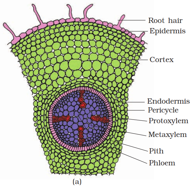
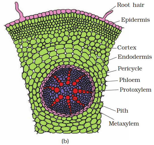
Draw illustrations to bring out the anatomical difference between: Monocot root and Dicot root.
|
S.No.
|
Monocot Root
|
Dicot Root
|
|
1.
|
Cortex is very wide.
|
Cortex is comparatively narrow.
|
|
2.
|
Outer cortex is usually differentiated into exodermis.
|
Outer cortex is usually not differentiated into exodermis.
|
|
3.
|
Endodermis contains prominent casparian strips only in young roots.
|
Endodermis contains prominant casparian strips.
|
|
4.
|
Xylem and phloem bundles are many in number 8 or more.
|
Xylem and phloem bundles vary in number from 2-6.
|
|
5.
|
Metaxylem vessels are oval or rounded.
|
Metaxylem vessels generally angular.
|
|
6.
|
No secondary growth.
|
Secondary growth takes place.
|
|
7.
|
Pith is large and well-developed.
|
Pith is small or absent.
|
|
|
 |
 |
Generate a complete, print-ready paper with questions like this in minutes — across 16+ boards, with answer keys.
|
A.
|
Resting potential
|
i.
|
Chemicals involved in the transmission of impulses at synapses.
|
|
B.
|
Nerve impulse
|
ii.
|
Gap between the pre synaptic and post synaptic neurons.
|
|
C.
|
Synaptic cleft
|
iii.
|
Electrical potential difference across the resting neural membrane.
|
|
D.
|
Neurotransmitters
|
iv.
|
An electrical wave like response of a neuron to a stimulation.
|